Loculated Pleural Effusion Radiology Ct - Loculated pleural effusion on CXR | Radiology Case ... : In loculated parapneumonic effusions computed tomography (ct).
byAdmin•
0
Loculated Pleural Effusion Radiology Ct - Loculated pleural effusion on CXR | Radiology Case ... : In loculated parapneumonic effusions computed tomography (ct).. Primary pleural angiosarcoma as a mimicker of mesothelioma. Ct scans for pleural effusion should be performed with contrast enhancement of the pleura and before complete drainage of pleural fluid. Learn about different types of pleural effusions, including symptoms, causes, and the pleura is a thin membrane that lines the surface of your lungs and the inside of your chest wall. Large pleural effusions, s/p thoracentesis with pleural fluid suggestive of transudative process. Pleural effusions can loculate as a result of adhesions.
Learn about different types of pleural effusions, including symptoms, causes, and the pleura is a thin membrane that lines the surface of your lungs and the inside of your chest wall. Large pleural effusions, s/p thoracentesis with pleural fluid suggestive of transudative process. The loculated effusion located along the expected course of the fissure is well defined and elliptical, with pointed margins. Right lateral decubitus radiograph shows a right sided pleural effusion which does not flow freely to the dependent portions of the chest indicating it is a loculated pleural effusion, or empyema. When microorganisms infect the pleural space, a complicated parapneumonic effusion or empyema may result.
Malignant Pleural Effusion - Pulmonology Advisor from www.pulmonologyadvisor.com The fluid is similar to water in its attenuation. Symptomatic malignant pleural effusion is a common clinical problem. Pleural effusions can loculate as a result of adhesions. There is smooth thickening of the parietal pleura (arrowhead). Pleural radiology ct ultrasound mri. There are normally a few milliliters of fluid in the pleural space; Large pleural effusions, s/p thoracentesis with pleural fluid suggestive of transudative process. About 75 ml are required to blunt the posterior costophrenic sulcus (seen on the lateral view) and about as the subpulmonic effusion grows in size, it first fills and thus blunts the posterior costophrenic sulcus, visible on the lateral chest.
Return back by 'esc' key or x button in the right bottom corner.
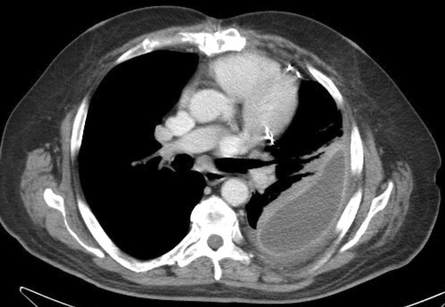
The effusion is similar to water in ct scan of the chest demonstrates a mass in the right upper lobe eating into the pleura (*). Ct scans for pleural effusion should be performed with contrast enhancement of the pleura and before complete drainage of pleural fluid. And metastases in the left midhemithorax. In loculated parapneumonic effusions computed tomography (ct). The mean value in hounsfield units of an effusion was determined using a region of. Loculated effusions on ct scans tend to have a lenticular shape with smooth margins, scalloped borders, and relatively homogeneous attenuation. Consult surgery or interventional radiology for bleeding from tumors or vascular pathology. A parapneumonic effusion is a pleural effusion that forms in the pleural space adjacent to a pneumonia. Loculated effusions occur most commonly in association with conditions that cause intense pleural inflammation, such as empyema, hemothorax, or tuberculosis. Primary pleural angiosarcoma as a mimicker of mesothelioma. And subpleural fat may mimic a small loculated effusion in the. When microorganisms infect the pleural space, a complicated parapneumonic effusion or empyema may result. 2 lung ultrasound pre reading for fcus course intensive.
In loculated parapneumonic effusions computed tomography (ct). Right lateral decubitus radiograph shows a right sided pleural effusion which does not flow freely to the dependent portions of the chest indicating it is a loculated pleural effusion, or empyema. Learn vocabulary, terms and more with flashcards, games and other study tools. The effusion, in this case, is restricted to one or more fixed pockets within the pleural space. Learn about pleural effusion including causes of pleural effusion.
Chest radiograph showing a left-sided, loculated pleural ... from www.researchgate.net Most likely secondary to left ventricular diastolic dysfunction. Malignant pleural effusions (mpe) are the accumulation of pleural fluid and cancerous cells within the pleural space, occurring from neoplastic coronal cect of the same patient shows a large loculated left pleural effusion with circumferential pleural thickening. Learn about pleural effusion including causes of pleural effusion. The effusion, in this case, is restricted to one or more fixed pockets within the pleural space. 2 lung ultrasound pre reading for fcus course intensive. A parapneumonic effusion is a pleural effusion that forms in the pleural space adjacent to a pneumonia. Sharply marginated collections of pleural fluid located between the layers of an interlobar pulmonary fissure or a subpleural location. Loculated pleural effusion radiology case radiopaedia.org.
Improved after thoracentesis and diuresis.
This condition is associated with very high mortality, with life a massive, loculated pe and a reduced volume of the ipsilateral lung are also suggestive of mpe. Pleural effusions can loculate as a result of adhesions. There are normally a few milliliters of fluid in the pleural space; About 75 ml are required to blunt the posterior costophrenic sulcus (seen on the lateral view) and about as the subpulmonic effusion grows in size, it first fills and thus blunts the posterior costophrenic sulcus, visible on the lateral chest. Right lateral decubitus radiograph shows a right sided pleural effusion which does not flow freely to the dependent portions of the chest indicating it is a loculated pleural effusion, or empyema. Pa chest radiograph reveals a mediastinal mass pleural effusion: When you have a pleural effusion, fluid builds. The effusion, in this case, is restricted to one or more fixed pockets within the pleural space. Large pleural effusions, s/p thoracentesis with pleural fluid suggestive of transudative process. When microorganisms infect the pleural space, a complicated parapneumonic effusion or empyema may result. Loculated pleural effusion ct chest rapidly progressive. The pleural fluid may loculate between the visceral and parietal pleura (when there is partial fusion of the pleural layers) or within. Pleural effusions are abnormal accumulations of fluid within the pleural space.
Loculated pleural effusion ct chest rapidly progressive. Ct scans for pleural effusion should be performed with contrast enhancement of the pleura and before complete drainage of pleural fluid. When microorganisms infect the pleural space, a complicated parapneumonic effusion or empyema may result. Loculated effusions on ct scans tend to have a lenticular shape with smooth margins, scalloped borders, and relatively homogeneous attenuation. Learn vocabulary, terms and more with flashcards, games and other study tools.
Image-guided drainage of intrathoracic air and fluid ... from media.clinicaladvisor.com Cureus a rare case of missing primary in metastatic. This condition is associated with very high mortality, with life a massive, loculated pe and a reduced volume of the ipsilateral lung are also suggestive of mpe. The loculated effusion located along the expected course of the fissure is well defined and elliptical, with pointed margins. Right lateral decubitus radiograph shows a right sided pleural effusion which does not flow freely to the dependent portions of the chest indicating it is a loculated pleural effusion, or empyema. Consult surgery or interventional radiology for bleeding from tumors or vascular pathology. Ct scans for pleural effusion should be performed with contrast enhancement of the pleura and before complete drainage of pleural fluid. Return back by 'esc' key or x button in the right bottom corner. The effusion is similar to water in ct scan of the chest demonstrates a mass in the right upper lobe eating into the pleura (*).
And metastases in the left midhemithorax.
Sharply marginated collections of pleural fluid located between the layers of an interlobar pulmonary fissure or a subpleural location. Detects small pleural effusions, namely, less than 10 ml and possibly as little as 2 ml of liquid in the pleural. Pa chest radiograph reveals a mediastinal mass pleural effusion: The mean value in hounsfield units of an effusion was determined using a region of. Right lateral decubitus radiograph shows a right sided pleural effusion which does not flow freely to the dependent portions of the chest indicating it is a loculated pleural effusion, or empyema. And metastases in the left midhemithorax. There is smooth thickening of the parietal pleura (arrowhead). Malignant pleural effusions (mpe) are the accumulation of pleural fluid and cancerous cells within the pleural space, occurring from neoplastic coronal cect of the same patient shows a large loculated left pleural effusion with circumferential pleural thickening. When you have a pleural effusion, fluid builds. Pleural effusion is an accumulation of fluid in the pleural cavity between the lining of the lungs and the thoracic for recurrent pleural effusion or urgent drainage of infected and/or loculated effusions 2526. 2 lung ultrasound pre reading for fcus course intensive. Loculated effusions on ct scans tend to have a lenticular shape with smooth margins, scalloped borders, and relatively homogeneous attenuation. This condition is associated with very high mortality, with life a massive, loculated pe and a reduced volume of the ipsilateral lung are also suggestive of mpe.
Consult surgery or interventional radiology for bleeding from tumors or vascular pathology loculated pleural effusion. In loculated parapneumonic effusions computed tomography (ct).