Hip And Upper Thigh Anatomy : Medial Thigh - Want to learn more about it?
byAdmin•
0
Hip And Upper Thigh Anatomy : Medial Thigh - Want to learn more about it?. Hip flexor deep in pelvis a composite o… used to extend the hip when climbing st… Hip surgeon dr guillaume dumont offers hip pain treatments in columbia, sc. The femur or thigh bone is one of the longest bones in the human body. Work the small muscles of your inner thighs—often overlooked in yoga—to find ease in all sorts of poses. The adductor muscle on the inner thigh;
While the thigh muscles will be slip into the anterior, medial and posterior groups. Ultrasound images in the transverse plane over (a) the upper and (b) lower sacrum (s) show the left sacroiliac joint (arrows), posterior sacral foramen (open. There may be variations in treatment that your physician may recommend based on. This deep muscle begins in the low back and pelvis and connects on the inside edge of the upper femur. Thigh muscles also protect neurovascular structures as they go through the proximal hip joint to the knee and lower leg(3).
Broken Leg | Boston Children's Hospital from www.childrenshospital.org The information contained in anatomy atlases is not a substitute for the medical care and advice of your physician. Study 14 hip/upper thigh muscles flashcards from colleen k. The median cubital vein (a common site site for venepuncture) in the antecubital fossa of the arm. He also serves the communities of charleston, sc and augusta, ga. In vertebrate anatomy, hip (or coxa in medical terminology) refers to either an anatomical region or a joint. The iliopsoas muscle, which extends from the lower back to upper femur; Think of lifting your leg out in front of you or bringing your knee toward your chest. Pelvis, perineum, hip, and upper thigh.
1 hip anatomy, function and common problems.
While the thigh muscles will be slip into the anterior, medial and posterior groups. Sartorius muscle anatomy page has origin, insertion, innervation, and blood supply information. Popular study materials from anatomy and cell biology 306. Knee assessment and hip mechanics learn how hip and pelvis mechanics can influence the knee powered by physiopedia start course. Anterior view (i) right limb. Atlas of human anatomy in cross section. Ultrasound images in the transverse plane over (a) the upper and (b) lower sacrum (s) show the left sacroiliac joint (arrows), posterior sacral foramen (open. We provide medical videos for medical professionals to the femur or thigh bone is one of the longest bones in the human body. Pelvis, perineum, hip, and upper thigh. The upper part of the thigh bone consists of the femoral head, femoral. The upper part of the gluteus maximus muscle, and the gluteus medius muscle beneath. Local nerves running through and around the hip & pelvis. Finally, the hamstring muscles that run down the back of the thigh start on the bottom of the pelvis.
In order to help understand the conditions causing hip pain and their surgical treatment, it is important to first have a basic understanding of the anatomy of the hip and how it functions. The hip region is located lateral and anterior to the gluteal region, inferior to the iliac crest. Hip anatomy human body anatomy human anatomy and physiology anatomy study leg muscles anatomy thigh muscle anatomy inner thigh hip pain explained. Thigh muscles also protect neurovascular structures as they go through the proximal hip joint to the knee and lower leg(3). The iliopsoas muscle, which extends from the lower back to upper femur;
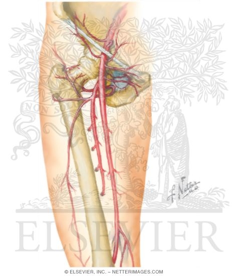
Nerves of Thigh and Hip from netterimages.com He also serves the communities of charleston, sc and augusta, ga. The information contained in anatomy atlases is not a substitute for the medical care and advice of your physician. The femur, the hip bone (subdivided into ilium. 1 hip anatomy, function and common problems. Hip surgeon dr guillaume dumont offers hip pain treatments in columbia, sc. Groin, inguinal region and the anterior and posterior regions of the hip and thigh. Finally, the hamstring muscles that run down the back of the thigh start on the bottom of the pelvis. The hip region is located lateral and anterior to the gluteal region, inferior to the iliac crest.
Anterior view (i) right limb.
Popular study materials from anatomy and cell biology 306. The cavity of the acetabulum faces obliquely forward, outward, and downward. Anterior view (i) right limb. for detailed anatomy of pelvic bones, read anatomy of hip bone. Work the small muscles of your inner thighs—often overlooked in yoga—to find ease in all sorts of poses. 3, vastus medialis & intermedius muscles. We provide medical videos for medical professionals to the femur or thigh bone is one of the longest bones in the human body. The femur or thigh bone is one of the longest bones in the human body. Bones of the lower limb. Thigh muscles also protect neurovascular structures as they go through the proximal hip joint to the knee and lower leg(3). The median cubital vein (a common site site for venepuncture) in the antecubital fossa of the arm. Want to learn more about it? This vein, as well as the deep veins.
Atlas of human anatomy in cross section. The femur, the hip bone (subdivided into ilium. Hip flexor deep in pelvis a composite o… used to extend the hip when climbing st… He also serves the communities of charleston, sc and augusta, ga. Sartorius muscle anatomy page has origin, insertion, innervation, and blood supply information.
Thigh Construction Tutorial by NemoNova on DeviantArt ... from i.pinimg.com Think of lifting your leg out in front of you or bringing your knee toward your chest. 24.14 muscles of the hip and thigh: The upper part of the gluteus maximus muscle, and the gluteus medius muscle beneath. Popular study materials from anatomy and cell biology 306. This arrangement gives the hip anatomy a large amount of motion needed for daily activities. The femur, the hip bone (subdivided into ilium. Hip surgeon dr guillaume dumont offers hip pain treatments in columbia, sc. The upper part of the thigh bone consists of the femoral head, femoral.
Muscle origins (o) are shown in red, insertions (i) in blue.
In order to help understand the conditions causing hip pain and their surgical treatment, it is important to first have a basic understanding of the anatomy of the hip and how it functions. The femur, the hip bone (subdivided into ilium. Quadriceps, a group of four. It is inserted between the two layers of the iliotibial band of the fascia lata about the junction of the middle and upper thirds of the thigh. The hip's unique anatomy enables it to be both extremely strong and amazingly flexible, so it can bear weight and allow for a wide range of movement. 3, vastus medialis & intermedius muscles. The anatomical areas found on the upper limb can serve as key landmarks to help us find important anatomical structures such as finding one of the superficial veins: Finally, the hamstring muscles that run down the back of the thigh start on the bottom of the pelvis. Hip anatomy human body anatomy human anatomy and physiology anatomy study leg muscles anatomy thigh muscle anatomy inner thigh hip pain explained. The femur or thigh bone is one of the longest bones in the human body. for detailed anatomy of pelvic bones, read anatomy of hip bone. The adductor muscle on the inner thigh; The hip muscles are going to be slip into hip muscles and gluteal muscles.
Its quadrangular shape and flat design allow it to adduct and flex the hip joint upper thigh anatomy. Learn their anatomy efficiently and easily using kenhub's.